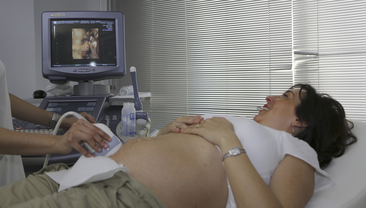
Handbook on 3D and 4D Ultrasounds:Obtaining ultrasound images of your baby is a cherished aspect of pregnancy, especially with the advancement of 3D and 4D ultrasounds, which provide remarkably detailed and realistic pictures. However, it’s crucial to note that experts strongly advise against opting for ‘keepsake’ ultrasounds conducted in non-medical environments due to potential safety concerns and lack of proper medical oversight. Your baby’s health and safety should always be the top priority.
Following the challenges of early pregnancy, experiencing an ultrasound and glimpsing images of your baby can be a significant delight. While 2-D ultrasounds remain the norm for routine check-ups, the availability of 3D and 4D ultrasounds is on the rise. This advanced technology can provide further insights and sharper perspectives of your growing baby, enhancing the bonding experience for expectant parents.
What is 3D Ultrasound?
A 3D ultrasound is used during pregnancy to observe the baby and the uterus, employing high-frequency sound waves akin to 2D ultrasounds. While 2D ultrasounds produce cross-sectional views of the baby in black, white, and gray, 3D ultrasounds offer more detailed, three-dimensional images by capturing numerous 2D images and merging them.
This method provides clearer views of the baby’s face or other organs, resembling real photographs rather than traditional ultrasound images. Although standard prenatal care mainly relies on 2D imaging, 3D ultrasounds are advantageous for detecting specific anomalies, such as clubfoot or cleft lip and palate, which may be difficult to discern with conventional 2D ultrasounds. In roughly 10% of cases, specific perspectives are inaccessible with 2D ultrasounds, making 3D ultrasounds indispensable for comprehensive prenatal assessment.
What is 4D Ultrasound?
A 4D ultrasound is a 3D ultrasound in motion, where a sequence of 3D images is compiled to create a low-resolution video. It allows expectant parents to witness their baby’s movements, such as sucking their thumb, opening and closing their eyes, and even smiling. Although 4D ultrasounds are not commonly employed for medical purposes and are not routinely conducted by doctors, they may be utilized in exceptional circumstances to observe fetal movements and assess brain development.
Is 3D Ultrasound Safe?
Yes, 3D ultrasound, like 2D ultrasound, is considered safe by trained medical professionals. According to both the March of Dimes and the American College of Obstetricians and Gynecologists (ACOG), there is no evidence linking ultrasound to harmful effects on a developing fetus, such as congenital disabilities, childhood cancer, or developmental issues later in life. However, some experts advocate for caution and recommend limiting exposure to ultrasound, adhering to the principle of “As Low As Reasonably Achievable” (ALARA).
This principle guides ultrasound technicians in minimizing prenatal exposure to ultrasound waves. While ultrasound is generally considered safe with low risks, prolonged exposure or operation by untrained individuals may pose increased risks. FDA warns that ultrasound waves can cause slight tissue heating and produce small gas pockets in body fluids or tissues, with uncertain long-term effects. For this reason, the FDA and other experts discourage the use of keepsake ultrasounds – 3D and 4D ultrasounds performed for recreational rather than medical purposes.
Should I Get a 3D or 4D Ultrasound?
When deliberating whether to opt for a 3D or 4D ultrasound during pregnancy, it’s imperative to assert your decision-making authority, considering both medical necessity and personal preference, under the expert guidance of your healthcare provider. While these cutting-edge imaging techniques can unveil intricate details of your baby’s development, they are typically reserved for situations necessitating additional insights beyond those offered by a conventional 2D scan. Hence, engaging in a proactive dialogue with your healthcare provider is paramount to ascertain if a 3D or 4D ultrasound is warranted in your unique circumstances.
During this proactive discourse, your healthcare provider will meticulously evaluate your medical requirements and weigh any potential benefits or risks of these advanced imaging modalities. They will furnish comprehensive guidance on the appropriateness of a 3D or 4D ultrasound and elucidate any preparatory measures or considerations requisite for the procedure.
It’s pivotal to recognize that while 3D and 4D ultrasounds can furnish captivating imagery and dynamic videos of your baby’s movements, they may not invariably constitute a medical imperative and might not be encompassed by insurance coverage unless deemed indispensable by your healthcare provider. Consequently, thoroughly comprehending the potential financial implications and insurance coverage beforehand is essential.
Ultimately, your active participation and collaboration with your healthcare provider, amalgamating medical necessities and personal preferences, should encourage you to pursue a 3D or 4D ultrasound. Through this assertive partnership, you can make a well-informed determination that optimally aligns with your needs and desires during this momentous phase of your pregnancy.
What Week is Best for a 3D Ultrasound?
The recommended timing for a 3D ultrasound is mainly between 18 and 22 weeks of pregnancy, often coinciding with the mid-pregnancy ultrasound. This period is optimal for obtaining clear images of the baby’s features. However, as the pregnancy progresses and the baby shifts into a head-down position, obtaining good 3D images becomes more challenging. Nevertheless, it’s still possible to capture beautiful face pictures later in pregnancy under the right conditions, such as the baby being in the correct position and with sufficient amniotic fluid. Despite the optimal window being between 18 and 22 weeks, if your healthcare provider believes a 3D ultrasound could provide valuable information, they may recommend it at any stage of pregnancy. Additionally, suppose you undergo an ultrasound in the first trimester for pregnancy confirmation or dating. In that case, you can inquire about a 3D image, although the baby’s features may not be as defined at that early stage.
How Much Does a 3D Ultrasound Cost?
The cost of a 3D ultrasound varies depending on where you go and if it’s deemed medically necessary. Insurance usually covers 2D ultrasounds but might only cover 3D if medically needed. Doctors can’t charge for non-medical 3D images but might provide them during a 2D scan or charge the insurer. It’s wise to discuss costs with your insurer and provider beforehand. Generally, a 3D ultrasound at a medical office can cost $1,000 to $2,000 if paid out of pocket. Keepsake 3D and 4D ultrasounds from specialized places may range from $100 to $400, but experts advise against unnecessary ultrasounds.

